Hello, Sign in
My Account
My Account
New customer? Start here


| Catalog # | Availability | Size | Quantity | Unit Price | Save For Later Wish List | |
|---|---|---|---|---|---|---|
| ABG1011-100 | 3 days | 100 µL | $499.00 |
Select product before adding to cart
|
||
| ABG1011-20 | 3 days | 20 µL | $149.00 |
Product Overview | |
| Name | KD-Validated Anti-MAP1LC3A Rabbit Monoclonal Antibody |
|---|---|
| Accession (Primary) | Q9H492 |
| Host Species | Rabbit |
| Clonality | Monoclonal |
| Isotype | Rabbit IgG |
| Clone Names | 23GB 1960 |
| Immunogen | |
| A synthesized peptide derived from human MAP1LC3A | |
| Species Reactivity | |
| Reacts With | Human, Mouse, Rat |
| Calculated MW | |
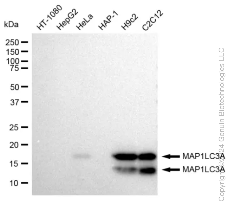
| Predicted, 14 kDa, observed, 14,16 kDa | |
| Storage Buffer | |
| Supplied in PBS (pH 7.4) containing 50% glycerol, and 0.02% sodium azide. | |
| Recommended Dilution | |
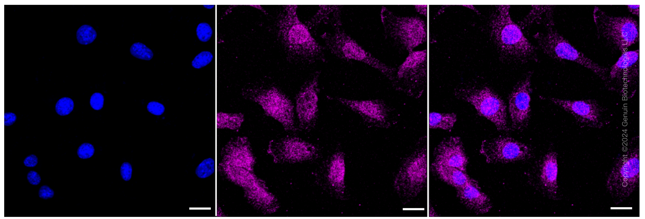
| Western Blotting (WB): 1:4,000-1:20,000 Immunocytochemistry (IC): 1:1,000 | |
Target Information: ( Q9H492 ) | |
| Gene Name | MAP1LC3A |
| Other Gene Names | |
| MAP1LC3A; Microtubule Associated Protein 1 Light Chain 3 Alpha; MAP1BLC3; MAP1ALC3; ATG8E; LC3A; LC3; Microtubule-Associated Proteins 1A/1B Light Chain 3A; Autophagy-Related Ubiquitin-Like Modifier LC3 A; MAP1 Light Chain 3-Like Protein 1; MAP1A/MAP1B Light Chain 3 A; MAP1A/MAP1B LC3 A; Microtubule-Associated Protein 1 Light Chain 3 Alpha; Microtubule-Associated Proteins 1A/1B Light Chain 3; Autophagy-Related Protein LC3 A; MAP1A/1B Light Chain 3 A | |
| About Gene Symbol | |
| MAP1LC3A (Microtubule Associated Protein 1 Light Chain 3 Alpha) is an autophagy regulatory protein involved in autophagy and ubiquitin-proteasome pathway. It is an important research target in cancer, cell signaling, and therapeutic target research and is widely studied to elucidate cellular mechanisms and identify therapeutic targets. | |

Thousands of laboratories across the world have published research that depended on the performance of antibodies from ADMEbio to advance their research.
Check out links to articles that cite our products in major peer-reviewed journals, organized by research category.
Thank you,
ADMEbio Team


ADMEbio welcomes feedback from our customers.
Thank you,
ADMEbio Team